INSIDE THE HEART
TAVI Assessment
Dr. John Jose E
Senior Interventional Cardiologist |
Structural Heart Specialist |
TAVI Specialist
 September 30, 2026
 10 minutes read
September 30, 2026
10 minutes read
Over the years, I have learned that the success of TAVI depends as much on the decisions we make before the procedure as on the procedure itself.
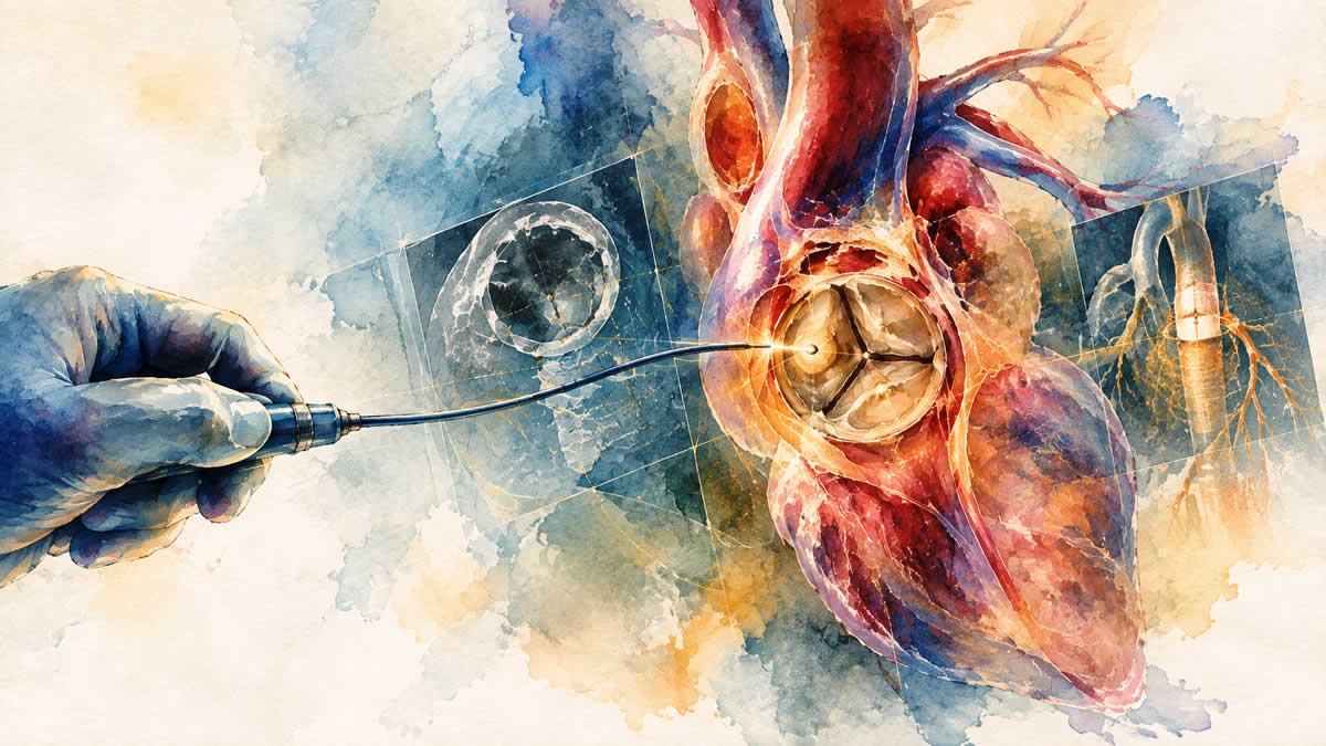
Before the first catheter enters, we need to understand the patient’s symptoms, study the heart carefully, assess the anatomy, determine the safest route to reach the heart and anticipate the challenges that may arise during the procedure.
Every scan, every measurement and every clinical detail contributes to the plan, from the valve we choose and the route we take to the way we position it and the challenges we prepare for.
For the patient and family, the catheterisation laboratory may be where they see the procedure happening.
For a structural heart specialist, the process begins much earlier.
It begins with assessment, imaging and planning.
A TAVI assessment begins with understanding why the patient has been referred to us.
Was it breathlessness?
A fall?
An episode of fainting?
Chest discomfort?
Or perhaps a gradual decline in activity that the family had simply accepted as part of ageing?
The clinical assessment helps us understand what the valve disease is actually doing to the patient’s life.
We take a detailed medical history, examine the patient and review investigations such as the echocardiogram and blood tests.
The echocardiogram tells us about the valve, the severity of narrowing and how the heart is responding to it.
But a report alone does not tell us everything.
We also need to understand the patient’s overall health, physical condition and other illnesses that may influence the safety and expected benefit of the procedure.
Kidney disease, lung disease, previous strokes, peripheral vascular disease, frailty and other medical problems can all change the risk profile.
This is why TAVI assessment is not based on a single test or number.
It is about putting many pieces together.
Severe aortic stenosis can develop gradually.
Patients often adapt their lives around their limitations without realising that they are doing so.
A father who once went for a morning walk may slowly stop going.
A mother who used to climb stairs comfortably may begin taking them one step at a time.
Someone who enjoyed meeting friends and going out with family may gradually start spending more time at home.
And often, the family thinks:
“Perhaps this is simply because they are getting older.”
I try to understand not only what a patient can do today, but what they have slowly stopped doing.
A patient may tell us:
“I am not really breathless.”
But when we ask whether they still walk to the shop, climb stairs, attend family functions or go out with friends, the answer may reveal a different story.
People can unknowingly adjust their lives around their limitations.
Breathlessness, chest discomfort, dizziness, fainting, unusual tiredness and a gradual reduction in activity can all be important signs of severe aortic stenosis.
Sometimes, what appears to be normal ageing is actually a heart valve that is quietly restricting a person’s life.
Understanding this is an important part of assessing how much the valve disease is affecting the patient’s daily life.
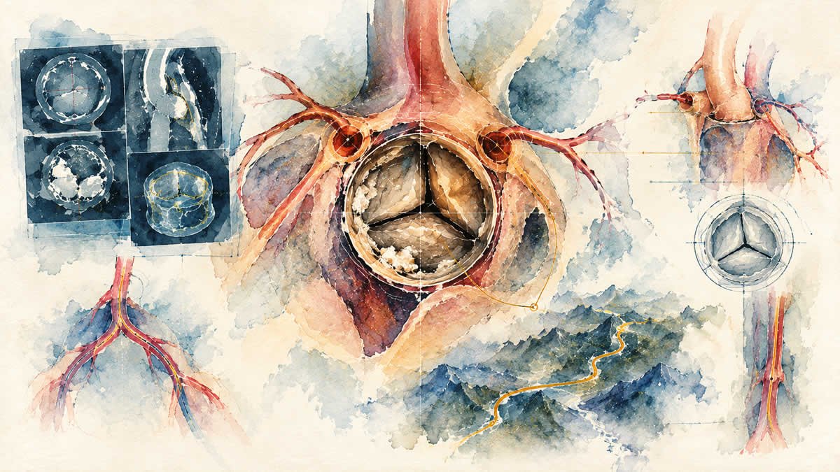
Once TAVI is being considered, the CT scan becomes one of the most important tools in planning the procedure.
When I look at a TAVI CT, I am not looking at it simply to confirm that the patient has aortic stenosis.
I am already thinking about what the images will mean for the procedure.
Where will the valve sit?
What is the relationship between the valve and the coronary arteries?
How much calcium is present, and where is it distributed?
What is the anatomy of the aortic root and surrounding structures?
Can we safely deliver the valve through the femoral arteries?
If not, what alternative route should we consider?
The annulus is carefully measured, and its dimensions and shape help guide valve selection and sizing.
We also consider whether the anatomy could increase the risk of coronary obstruction when the native valve leaflets are displaced during the procedure.
The CT gives us the detailed information needed to answer these questions.
In many ways, it is our roadmap.
For many patients, the femoral arteries in the groin are the preferred route for TAVI.
But especially in elderly patients, the arteries may not always be straightforward.
There may be severe calcification, tortuosity, previous stents, chronic blockages or other vascular problems.
These details matter.
The valve may be suitable.
The heart may be suitable.
But the route we use to reach the heart may still present a challenge.
The usual pathway is not always the safest pathway.
Advances in structural heart intervention, together with increasing experience with alternative access routes and newer technologies, have given us more options.
For a patient with challenging vascular anatomy, considering an alternative approach can sometimes make the difference between being able to offer a procedure and having very limited choices.
An unfavourable femoral route does not necessarily mean that TAVI is impossible.
It means that the access strategy needs to be reconsidered carefully.
Choosing a transcatheter valve is not simply a matter of selecting a particular brand or size.
The findings from imaging, including the anatomy of the aortic root, the extent and distribution of calcium and the dimensions of the annulus, all influence the choice.
We also consider how the implanted valve will relate to the coronary arteries and the heart’s conduction system.
The depth and position of the valve can influence the risk of conduction disturbances and the possible need for a pacemaker.
The goal is not simply to find a valve that fits.
It is to select the appropriate valve and size and determine the safest way to position and deploy it.
A few millimetres can matter.
Anatomy that may appear straightforward on one image can reveal important considerations when studied in detail.
The more carefully we understand the anatomy, the better prepared we are for what we may encounter.
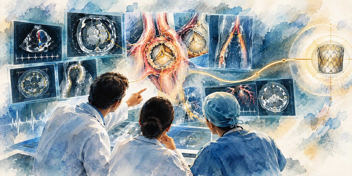
By this stage, we have gathered the clinical assessment, echocardiogram findings, CT information, vascular anatomy and an understanding of the patient’s overall health and procedural risk.
Now, these findings have to come together into a practical treatment strategy.
This is where the Heart Team discussion becomes important.
We ask:
Is TAVI appropriate for this patient?
What valve and size are most suitable?
What is the safest access route?
What anatomical or clinical challenges should we anticipate?
Would surgery offer a better option?
What are the expected benefits and risks of each approach?
The decision is not based simply on whether the procedure is technically possible.
We also consider the patient’s clinical risk, expected benefit, long-term considerations and, importantly, the patient’s own goals and preferences.
These questions have to be considered together rather than in isolation.
By the time the patient enters the catheterisation laboratory, the aim is to have a clear and individualised plan.
No procedure is completely predictable.
Even with excellent imaging and careful preparation, anatomy can sometimes behave differently from what we expected.
Good planning is not only about deciding what we intend to do.
It is also about anticipating what might happen if the expected pathway does not go as planned.
What if the vascular route proves more difficult?
What if the valve does not behave exactly as anticipated?
What if an unexpected anatomical challenge requires a change in strategy?
Preparation means thinking about these possibilities before they become problems.
The aim is not to expect complications.
It is to be prepared for them.
That is one of the important differences between performing a procedure and preparing for it.
For the patient and family, much of this planning may not be visible.
They may see the doctor reviewing scans on a screen.
They may hear discussions about valve sizes or access routes.
They may see members of different specialties discussing the case.
But behind those conversations is one objective:
to make the procedure as safe, precise and appropriate as possible for that individual patient.
The technology available to us today gives us remarkable possibilities.
But technology works best when it is combined with careful assessment, detailed imaging, experience and preparation.
When I walk into the catheterisation laboratory for a TAVI, I do not want the procedure to be the first time we are asking important questions.
The important decisions should already have been considered.
We should understand the anatomy.
We should know the strategy.
We should have anticipated the challenges.
And we should have a plan.
Of course, every procedure can bring something unexpected.
That is part of medicine.
But careful preparation allows us to approach those situations with greater confidence and clarity.
The procedure may take place in the catheterisation laboratory.
And often, the quality of that planning determines how confidently we can proceed when the time finally comes.
Because before we treat the valve, we must first understand the heart.
Every heart carries a story, and every story belongs to a person, a family and a life that matters.
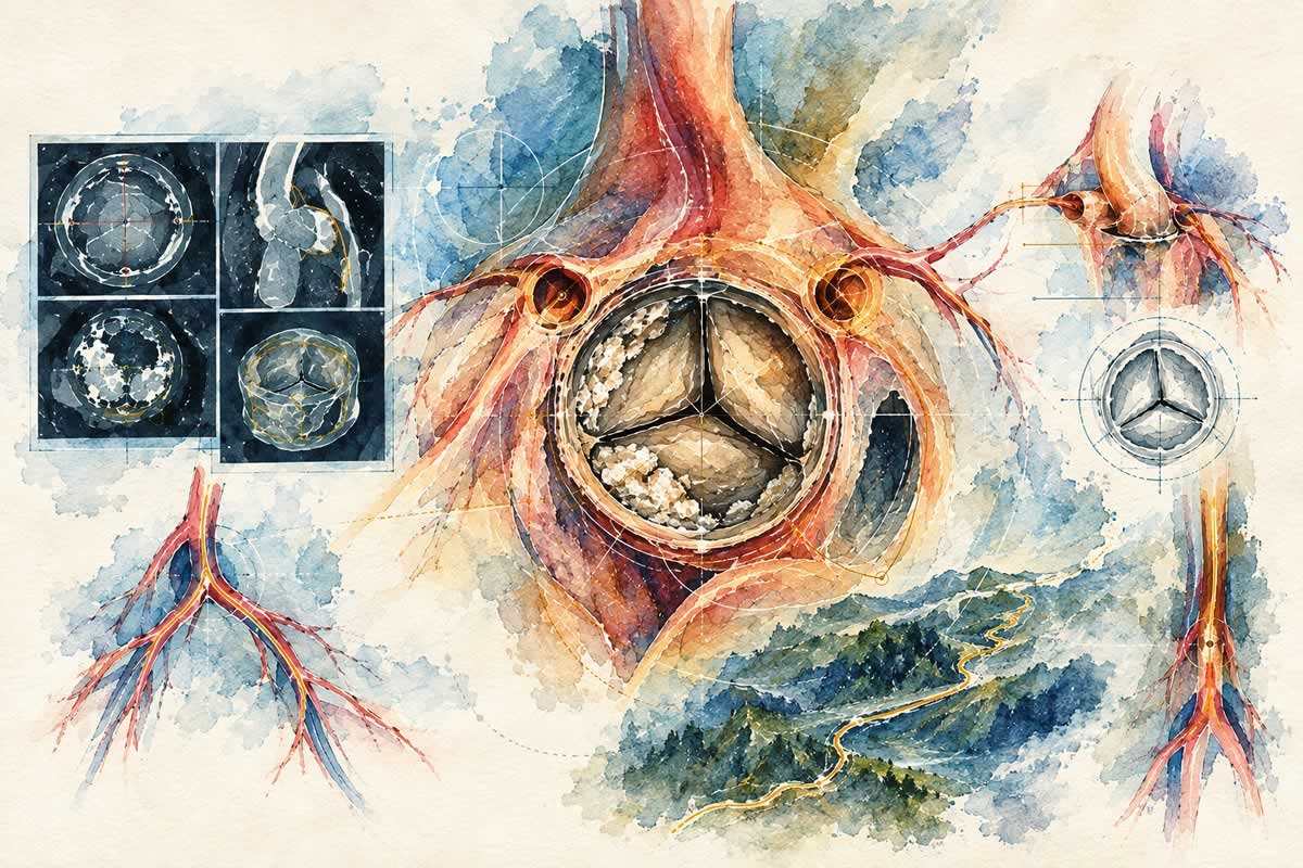
Once TAVI is being considered, the CT scan becomes one of the most important tools in planning the procedure.
When I look at a TAVI CT, I am not looking at it simply to confirm that the patient has aortic stenosis.
I am already thinking about what the images will mean for the procedure.
Where will the valve sit?
What is the relationship between the valve and the coronary arteries?
How much calcium is present, and where is it distributed?
What is the anatomy of the aortic root and surrounding structures?
Can we safely deliver the valve through the femoral arteries?
If not, what alternative route should we consider?
The annulus is carefully measured, and its dimensions and shape help guide valve selection and sizing.
We also consider whether the anatomy could increase the risk of coronary obstruction when the native valve leaflets are displaced during the procedure.
The CT gives us the detailed information needed to answer these questions.
In many ways, it is our roadmap.
For many patients, the femoral arteries in the groin are the preferred route for TAVI.
But especially in elderly patients, the arteries may not always be straightforward.
There may be severe calcification, tortuosity, previous stents, chronic blockages or other vascular problems.
These details matter.
The valve may be suitable.
The heart may be suitable.
But the route we use to reach the heart may still present a challenge.
The usual pathway is not always the safest pathway.
Advances in structural heart intervention, together with increasing experience with alternative access routes and newer technologies, have given us more options.
For a patient with challenging vascular anatomy, considering an alternative approach can sometimes make the difference between being able to offer a procedure and having very limited choices.
An unfavourable femoral route does not necessarily mean that TAVI is impossible.
It means that the access strategy needs to be reconsidered carefully.
Choosing a transcatheter valve is not simply a matter of selecting a particular brand or size.
The findings from imaging, including the anatomy of the aortic root, the extent and distribution of calcium and the dimensions of the annulus, all influence the choice.
We also consider how the implanted valve will relate to the coronary arteries and the heart’s conduction system.
The depth and position of the valve can influence the risk of conduction disturbances and the possible need for a pacemaker.
The goal is not simply to find a valve that fits.
It is to select the appropriate valve and size and determine the safest way to position and deploy it.
A few millimetres can matter.
Anatomy that may appear straightforward on one image can reveal important considerations when studied in detail.
The more carefully we understand the anatomy, the better prepared we are for what we may encounter.
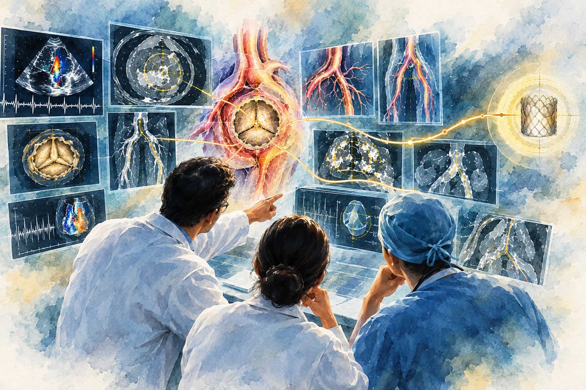
By this stage, we have gathered the clinical assessment, echocardiogram findings, CT information, vascular anatomy and an understanding of the patient’s overall health and procedural risk.
Now, these findings have to come together into a practical treatment strategy.
This is where the Heart Team discussion becomes important.
We ask:
Is TAVI appropriate for this patient?
What valve and size are most suitable?
What is the safest access route?
What anatomical or clinical challenges should we anticipate?
Would surgery offer a better option?
What are the expected benefits and risks of each approach?
The decision is not based simply on whether the procedure is technically possible.
We also consider the patient’s clinical risk, expected benefit, long-term considerations and, importantly, the patient’s own goals and preferences.
These questions have to be considered together rather than in isolation.
By the time the patient enters the catheterisation laboratory, the aim is to have a clear and individualised plan.
No procedure is completely predictable.
Even with excellent imaging and careful preparation, anatomy can sometimes behave differently from what we expected.
Good planning is not only about deciding what we intend to do.
It is also about anticipating what might happen if the expected pathway does not go as planned.
What if the vascular route proves more difficult?
What if the valve does not behave exactly as anticipated?
What if an unexpected anatomical challenge requires a change in strategy?
Preparation means thinking about these possibilities before they become problems.
The aim is not to expect complications.
It is to be prepared for them.
That is one of the important differences between performing a procedure and preparing for it.
For the patient and family, much of this planning may not be visible.
They may see the doctor reviewing scans on a screen.
They may hear discussions about valve sizes or access routes.
They may see members of different specialties discussing the case.
But behind those conversations is one objective:
to make the procedure as safe, precise and appropriate as possible for that individual patient.
The technology available to us today gives us remarkable possibilities.
But technology works best when it is combined with careful assessment, detailed imaging, experience and preparation.
When I walk into the catheterisation laboratory for a TAVI, I do not want the procedure to be the first time we are asking important questions.
The important decisions should already have been considered.
We should understand the anatomy.
We should know the strategy.
We should have anticipated the challenges.
And we should have a plan.
Of course, every procedure can bring something unexpected.
That is part of medicine.
But careful preparation allows us to approach those situations with greater confidence and clarity.
The procedure may take place in the catheterisation laboratory.
And often, the quality of that planning determines how confidently we can proceed when the time finally comes.
Because before we treat the valve, we must first understand the heart.
Every heart carries a story, and every story belongs to a person, a family and a life that matters.
Dr. John Jose E
Disclaimer:
The views expressed in my articles are my personal perspectives, shared for educational purposes and professional discussion. They are expressed in an individual capacity and do not necessarily represent the official views, policies, or positions of the organisations or institutions with which I am associated.
The information presented is intended for general awareness and education. It should not be considered a substitute for individual medical consultation, diagnosis or treatment. Medical decisions should be made in consultation with an appropriately qualified healthcare professional who can assess the patient’s individual circumstances. In a medical emergency, seek immediate medical assistance.